Olympus SoRa spinning disc
Room 214.00.09
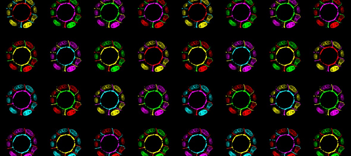
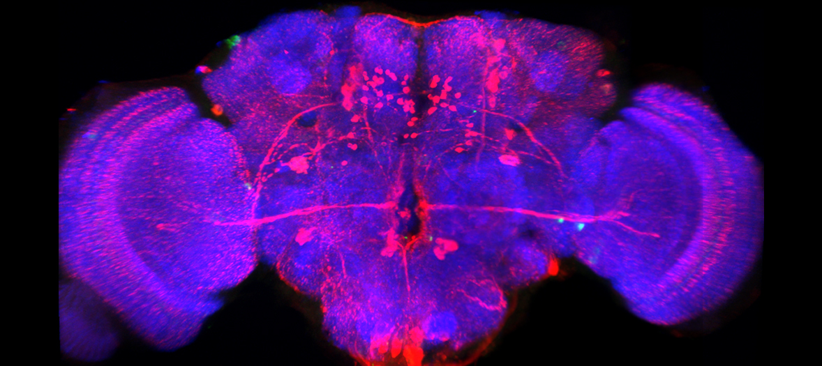
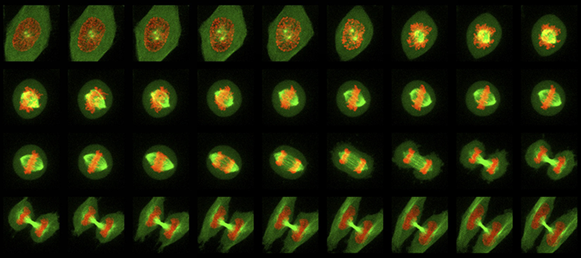
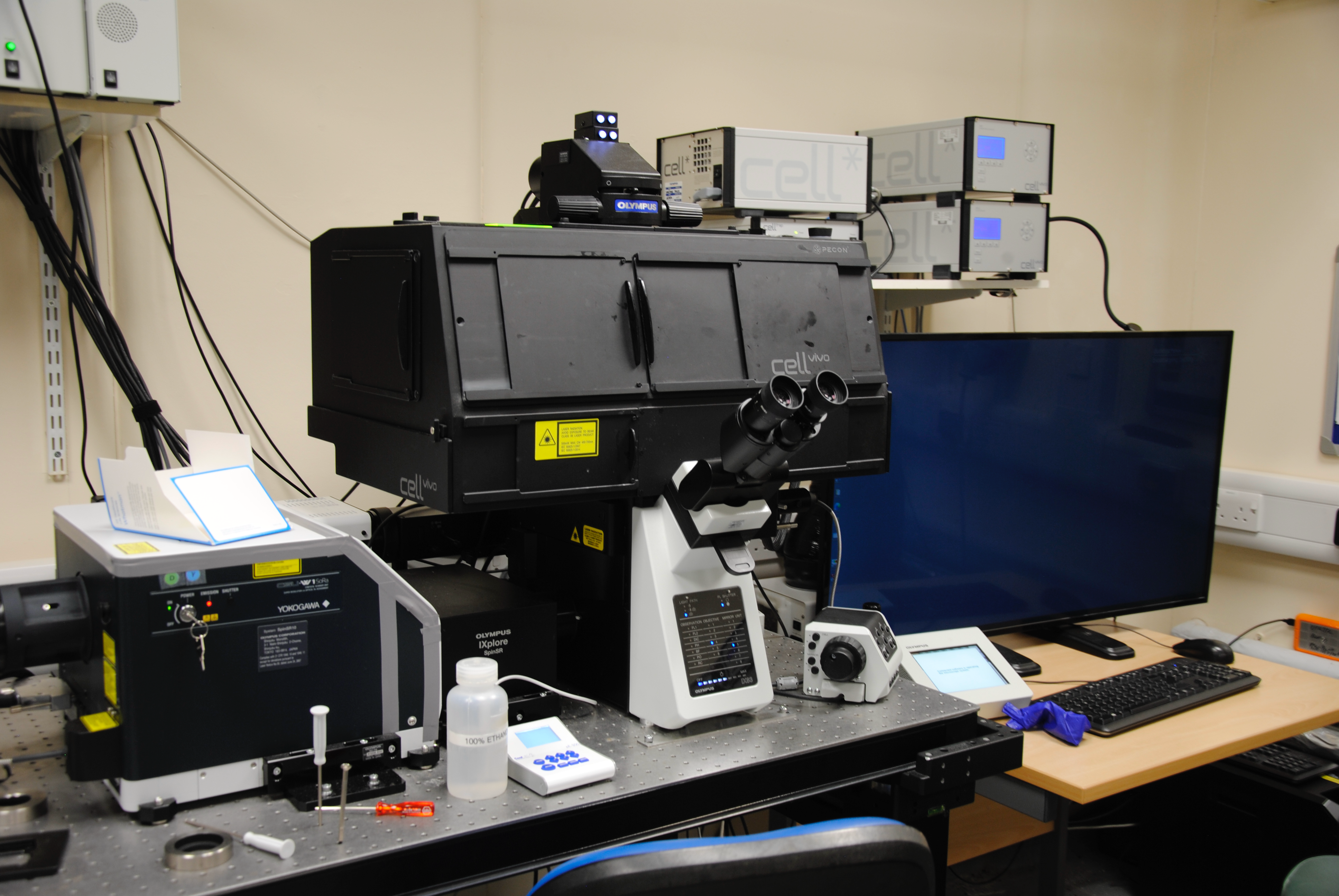
The Olympus SoRa spinning disc confocal is an inverted microscope for fast 3D super resolution imaging and prolonged cell viability in time-lapse experiments. In addition the system is fitted with z-peizo, ZDC focus, 405nm FRAP module and environmental chamber for super-resolution live cell imaging.
Applications:
- Fast, gentle imaging of live samples, possible to a resolution of ~120nm.
- Environmental chamber for time-lapse imaging at 37oC with 5% CO2.
- 405nm FRAP photomanipulation module
General specifications
- Olympus IX-83 inverted frame
- Yokogawa SoRa super-resolution spinning disc module
- Choice of cameras for either super-resolution imaging or maximised sensitivity
- Incubator system for live-cell imaging and temperature control
- 4 laser lines
- Freely selectable ROI for laser scanning, bleaching and photoactivation with 405nm laser line
- DIC optics
- Peizo stage insert
- ZDC focus
Olympus SoRa Laser Lines
| Laser | Excitation Lines | Suitable Dyes |
|---|---|---|
| Solid State | 405nm | Alexa 405, DAPI, Hoechst |
| Solid State | 488nm | Alexa 488, FITC, GFP, Cy2 |
| Solid State | 561nm | Alexa 546, TRITC, Cy3, DiI, propidium iodide, Texas red , mCherry |
| Solid State | 633nm | Alexa 633, Alexa 647, Cy5 |
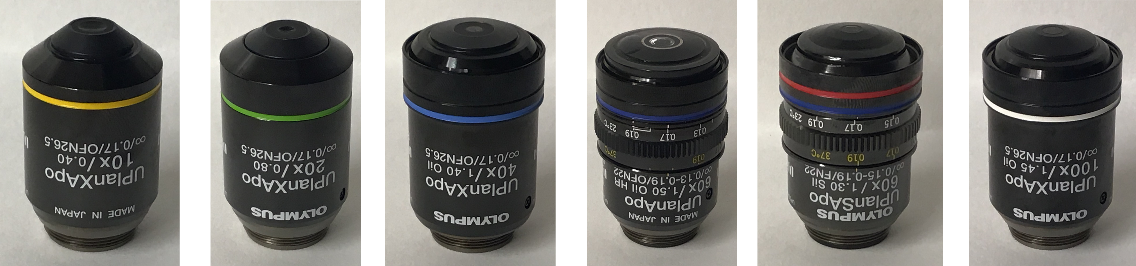
Objectives Available
Selected reading:
Spinning Disc Confocal Microscopy
- Azuma T, Kei T. (2015). Super-resolution spinning-disk confocal microscopy using optical photon reassignment. Optics Express, 23(11), 15003–15011. https://doi.org/10.1364/OE.23.015003 Nakano A. (2002). Spinning-disk confocal microscopy—a cutting-edge tool for imaging of membrane traffic. Cell Structure and Function, 27, 349–355.
- Graf R., Rietdorf J., Zimmermann T. (2005). Live cell spinning disk microscopy. Advances in Biochemical Engineering/Biotechnology, 95, 57–75.
Olympus SoRa Super-resolution
- Verth F., Fairn G.D. (2023). Super-Resolution Spinning-Disk Confocal Microscopy Using Optical Photon Reassignment (SoRa) to Visualize the Actin Cytoskeleton in Macrophages. Methods in Molecular Biology, 2692, 79–90.
- Verth F., et al. (2025). Super-resolution Spinning-Disk Confocal Microscopy Using Optical Photon Reassignment. Methods in Molecular Biology.
Fluorescence Recovery After Photobleaching (FRAP)
- Axelrod D., Koppel D.E., Schlessinger J., Elson E., Webb W.W. (1976). Mobility measurement by analysis of fluorescence photobleaching recovery kinetics. Biophysical Journal, 16, 1055–1069.
- Lippincott-Schwartz J., Snapp E., Kenworthy A. (2001). Studying protein dynamics in living cells. Nature Reviews Molecular Cell Biology, 2, 444–456.
- Sprague B.L., McNally J.G. (2005). FRAP analysis of binding: proper and fitting. Trends in Cell Biology, 15, 84–91.
DNA Damage by Laser Microirradiation (DNA Stripe Assay)
- Bekker-Jensen S., Lukas C., Melander F., et al. (2006). Spatial organization of the mammalian genome surveillance machinery in response to DNA strand breaks. Journal of Cell Biology, 173, 195–206.
- Lukas C., Falck J., Bartkova J., et al. (2003). Distinct spatiotemporal dynamics of mammalian checkpoint regulators induced by DNA damage. Nature Cell Biology, 5, 255–260.
- Kong X., Mohanty S.K., Stephens J., et al. (2009). Comparative analysis of different laser systems to study cellular responses to DNA damage in mammalian cells. Nucleic Acids Research, 37(9), e68.
General Live-cell Fluorescence Imaging
- Waters J.C. (2009). Accuracy and precision in quantitative fluorescence microscopy. Journal of Cell Biology, 185, 1135–1148.
- Waters J.C., Wittmann T. (2014). Concepts in quantitative fluorescence microscopy. Methods in Cell Biology, 123, 1–18.
Selected publications from the Olympus SoRa:
- PARP1-specific inhibitor displays PARP1-detrapping activity. Schützenhofer K, Laker E, Tsang K, Baretić D, Suskiewicz MJ, Zobel FFH, Simmons MDR, Chatrin C, Smith R, Ahel I. Cell Rep. 2026 May 26;45(5):117350. doi: 10.1016/j.celrep.2026.117350. Epub 2026 May 4. PMID: 42090288.
- Centrioles generate two scaffolds with distinct biophysical properties to build mitotic centrosomes. Wong SS, Monteiro JM, Chang CC, Peng M, Mohamad N, Steinacker TL, Xiao B, Saurya S, Wainman A, Raff JW. Sci Adv. 2025 Feb 7;11(6):eadq9549. doi: 10.1126/sciadv.adq9549. Epub 2025 Feb 7. PMID: 39919171; PMCID: PMC11804907.
- Mitochondrial origins of the pressure to sleep. Sarnataro R, Velasco CD, Monaco N, Kempf A, Miesenböck G. Nature. 2025 Sep;645(8081):722-728. doi: 10.1038/s41586-025-09261-y. Epub 2025 Jul 16. PMID: 40670797; PMCID: PMC12443607.
- Cell cycle dependent coordination of surface layer biogenesis in Caulobacter crescentus. Herdman M, Isbilir B, von Kügelgen A, Schulze U, Wainman A, Bharat TAM. Nat Commun. 2024 Apr 18;15(1):3355. doi: 10.1038/s41467-024-47529-5. PMID: 38637514; PMCID: PMC11026435.
- Perilipin membrane integration determines lipid droplet heterogeneity in differentiating adipocytes. Majchrzak M, Stojanović O, Ajjaji D, Ben M'barek K, Omrane M, Thiam AR, Klemm RW. Cell Rep. 2024 Apr 23;43(4):114093. doi: 10.1016/j.celrep.2024.114093. Epub 2024 Apr 10. PMID: 38602875.
- MPS1 localizes to end-on microtubule-attached kinetochores to promote microtubule release. Hayward D, Roberts E, Gruneberg U. Curr Biol. 2022 Dec 5;32(23):5200-5208.e8. doi: 10.1016/j.cub.2022.10.047. Epub 2022 Nov 16. PMID: 36395767.
Olympus SoRa
Training videos
Help Sheets

Spinning Disc Theory
