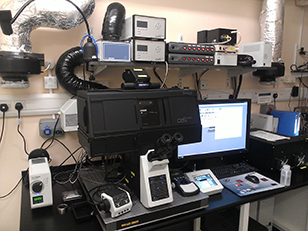
Olympus FV3000
Room 214.00.13
The FV3000 confocal is an inverted IX83 microscope equipped with sensitive GASP detectors.
Applications:
- Imaging fixed tissue culture cells tissue sections labelled with 3 or 4 fluorophores for cellular architecture, localisation or co-localisation studies.
- Imaging of thicker samples such as organoids, embryos etc.
- Programmable stage for imaging larger structures such as organ slices, or multiple points on a slide.
General Specifications
- FluoView Spectral FV3000 Laser Scanning Microscope based on a IX83 motorized inverted microscope
- 6 laser lines
- 2 standard PMT and 2 high sensitive GaAsP detectors.
- Scan speeds of up to 4 frames/second with 512 x 512 pixels
- xy, xz, xyz, xyzt, lambda, line scan and spot scan modes
FV3000 Laser Lines
| Laser | Excitation Lines | Suitable Dyes |
|---|---|---|
| Solid State | 405nm | Alexa 405, DAPI, Hoechst |
| Solid State | 455nm | CFP |
| Solid State | 488nm | Alexa 488, FITC, GFP, Cy2 |
| Solid State | 514nm | Alexa 514, YFP |
| Solid State | 561nm | Alexa 546, Alexa 568, TRITC, Cy3, DiI |
| Solid State | 594nm | Alexa 595, TRITC, Cy3, DiI, propidium iodide, Texas red, mCherry |
| Solid State | 640nm | Alexa 633, Alexa 647, Cy5 |
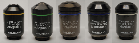
Objectives Available
Selected reading:
Selected publications from the FV3000:
Olympus FV3000
Training videos
Help Sheets