
FOCUSSED ION BEAM SCANNING ELECTRON MICROSCOPY (FIB-SEM)
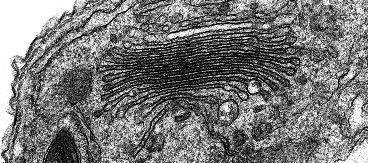
In volume SEM, either an in situ microtome (SBF-SEM/3View) or an ion beam (FIB-SEM) is used to remove a thin layer of resin and the block face is scanned with the electron beam, producing a TEM-like image; this automated process is then repeated until the set volume is reached. These techniques generate much larger volumes than TEM tomography, so are ideally suited to projects at the whole cell and tissue scale. SBF-SEM is excellent for studying tissue ultrastructure and cell populations in 3D, generating stacks upwards of 100 µm thick in a few days, with a lateral resolution of 10-20 nm and a z resolution of 70-200 nm. Complementary to this, FIB-SEM is the best option when isotropic resolution down to 5 nm3 is required, for instance to characterise the spatial organisation of synaptic vesicles in brain tissue or organelle contact sites in whole cells (see Kizilyaprak et al., Tissue and Cell, 2019).
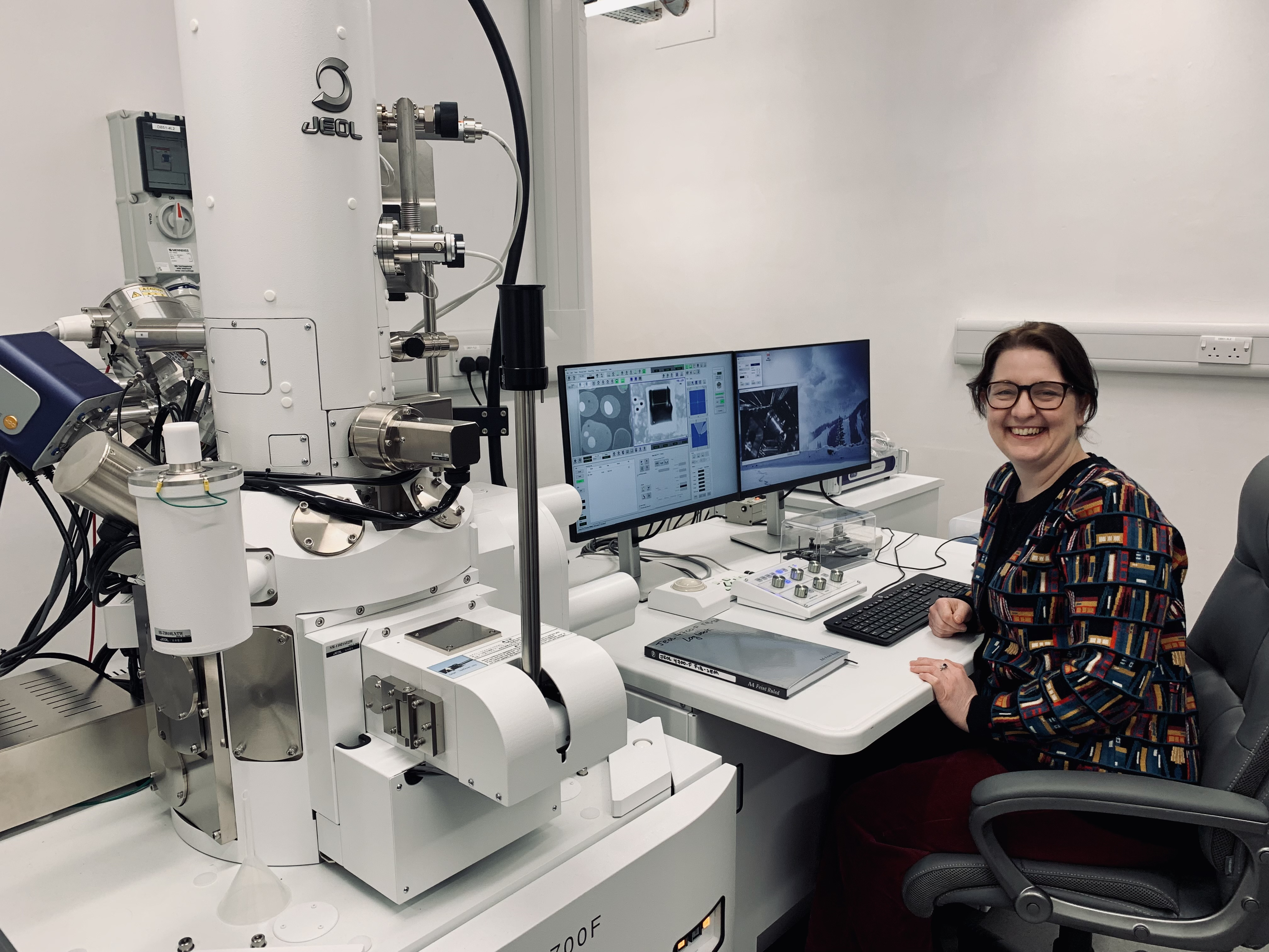
Our new JEOL 4700F will allow this powerful technique to be applied to biological EM projects for the first time at Oxford University. To learn more about FIB-SEM and how it could benefit your research, please contact Dr Charlotte Melia: charlotte.melia@path.ox.ac.uk.
Specifications (SEM column):
- Accelerating voltage: 0.1 to 30.0kV
- Image resolution (at optimum WD): 1.2nm (15kV, GB mode), 1.6nm (1kV, GB mode)
- Magnification: x20 to 1,000,000
- Probe current: 1pA to 300nA
- Detector: SE2 & BSE
- Specimen stage: Computerized 6-axis goniometer stage X: 50mm, Y: 50mm, Z: 1.5 to 40mm, R: 360°, T: -5 to 70°, FZ: -3.0 to +3.0mm
Specifications (FIB column):
- Accelerating voltage: 1 to 30kV
- Image resolution 4.0nm (30kV)
- Magnification: x50 to 1,000,000
- Probe current: 1 pA to 90 nA, 13 steps
Applications:
- 3D ultrastructural imaging of cells and tissue at isotropic resolution (eg: model synaptic architecture in 3D, model virus replication organelles in 3D)
- Correlative 3D light and electron microscopy
Recommended reading:
- Volume microscopy in biology: FIB-SEM tomography. Kizilyaprak C, Stierhof YD, Humbel BM. Tissue Cell. 2019 Apr;57:123-128. https://pubmed.ncbi.nlm.nih.gov/30385054/
- Correlative 3D imaging: CLSM and FIB-SEM tomography using high-pressure frozen, freeze-substituted biological samples. Lucas MS, Guenthert M, Gasser P, Lucas F, Wepf R. Methods Mol Biol. 2014;1117:593-616. https://pubmed.ncbi.nlm.nih.gov/24357381/
- Focused ion beams in biology. Narayan K, Subramaniam S. Nat Methods. 2015 Nov;12(11):1021-31. doi: https://pubmed.ncbi.nlm.nih.gov/26513553/
- A Complete Electron Microscopy Volume of the Brain of Adult Drosophila melanogaster. Zheng Z, Lauritzen JS, Perlman E, Robinson CG, Nichols M, Milkie D, Torrens O, Price J, Fisher CB, Sharifi N, Calle-Schuler SA, Kmecova L, Ali IJ, Karsh B, Trautman ET, Bogovic JA, Hanslovsky P, Jefferis GSXE, Kazhdan M, Khairy K, Saalfeld S, Fetter RD, Bock DD. Cell. 2018 Jul 26;174(3):730-743.e22. https://pubmed.ncbi.nlm.nih.gov/30033368/
- FIB-SEM as a Volume Electron Microscopy Approach to Study Cellular Architectures in SARS-CoV-2 and Other Viral Infections: A Practical Primer for a Virologist. Baena V, Conrad R, Friday P, Fitzgerald E, Kim T, Bernbaum J, Berensmann H, Harned A, Nagashima K, Narayan K. Viruses. 2021 Apr 2;13(4):611. https://pubmed.ncbi.nlm.nih.gov/33918371/
FIB-SEM
Resources
electron microscopy
electron microscopy
electron microscopy
