Olympus Fluoview FV3000
Room 214.00.13
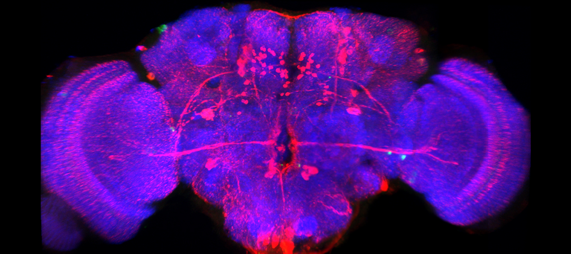
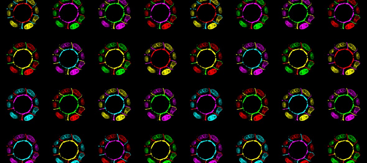
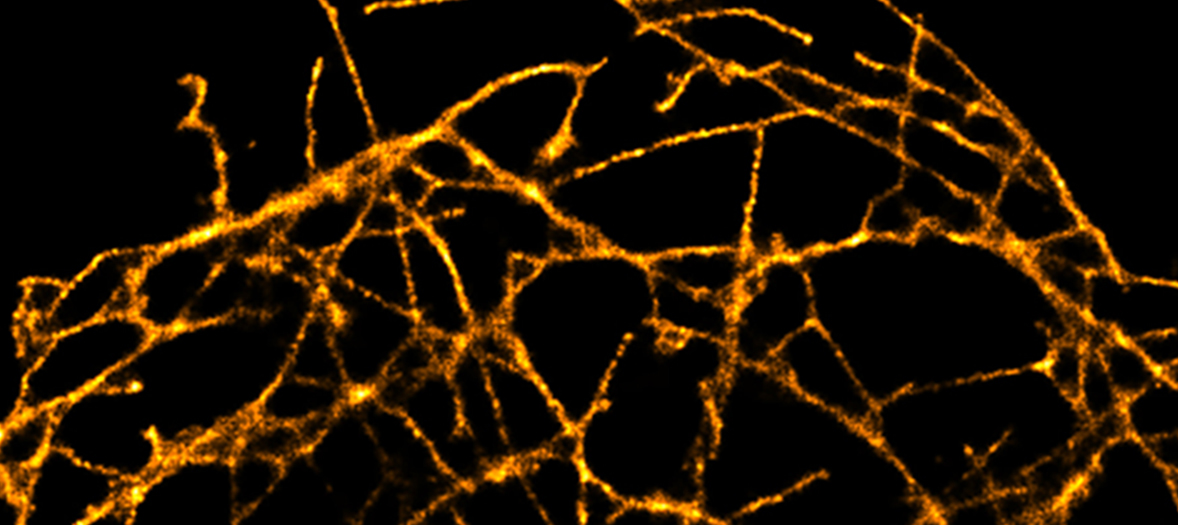
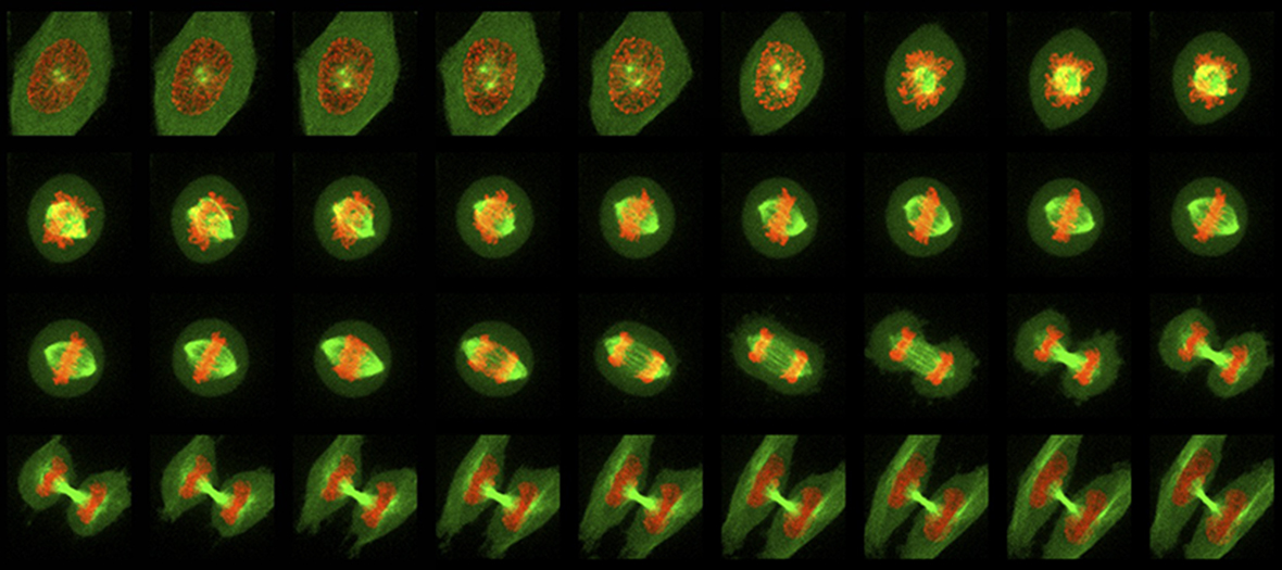
The FV3000 confocal is an inverted IX83 microscope equipped with sensitive GASP detectors. The motorized stage can be used for image stitching of large samples
Applications:
- Imaging fixed tissue culture cells tissue sections labelled with 3 or 4 fluorophores for cellular architecture, localisation or co-localisation studies.
- Imaging of thicker samples such as organoids, embryos etc.
- Programmable stage for imaging larger structures such as organ slices, or multiple points on a slide.
General Specifications
- FluoView Spectral FV3000 Laser Scanning Microscope based on a IX83 motorized inverted microscope
- 7 laser lines
- 2 standard PMT and 2 high sensitive GaAsP detectors.
- Scan speeds of up to 4 frames/second with 512 x 512 pixels
- xy, xz, xyz, xyzt, lambda, line scan and spot scan modes
FV3000 Laser Lines
| Laser | Excitation Lines | Suitable Dyes |
|---|---|---|
| Solid State | 405nm | Alexa 405, DAPI, Hoechst |
| Solid State | 445nm | CFP |
| 488nm | Alexa 488, FITC, GFP, Cy2 | |
| 514nm | Alexa 514, YFP | |
| Solid State | 551nm | Alexa 546, TRITC, Cy3, DiI, propidium iodide |
| Solid State | 594nm | Alexa 594, Texas red , mCherry |
| Solid State | 640nm | Alexa 633, Alexa 647, Cy5 |
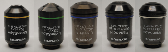
Objectives Available
Selected reading:
Selected publications from the FV3000:
Olympus FV3000

Training videos
Help Sheets