News
July 2019: Bioimaging Present at MMC conference
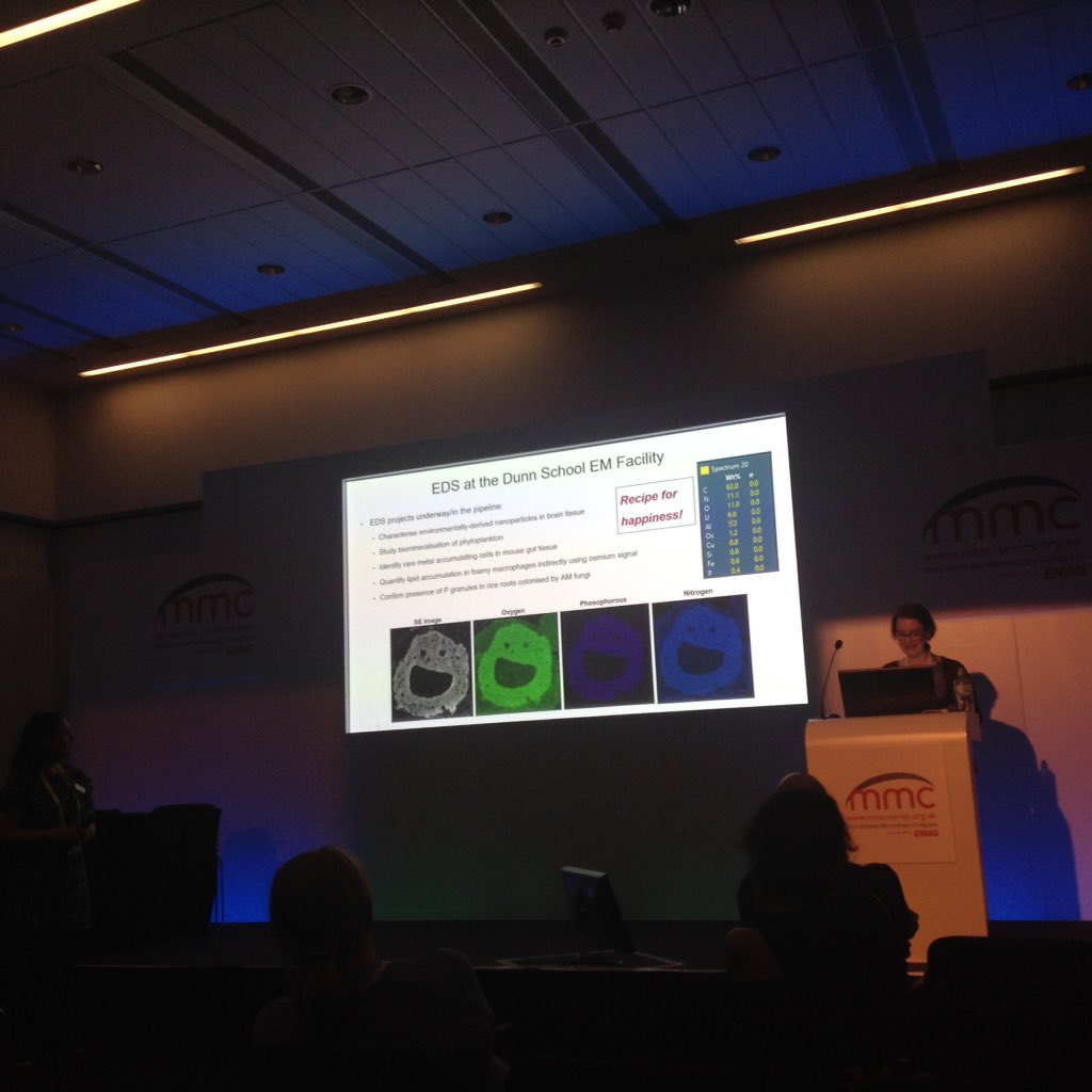
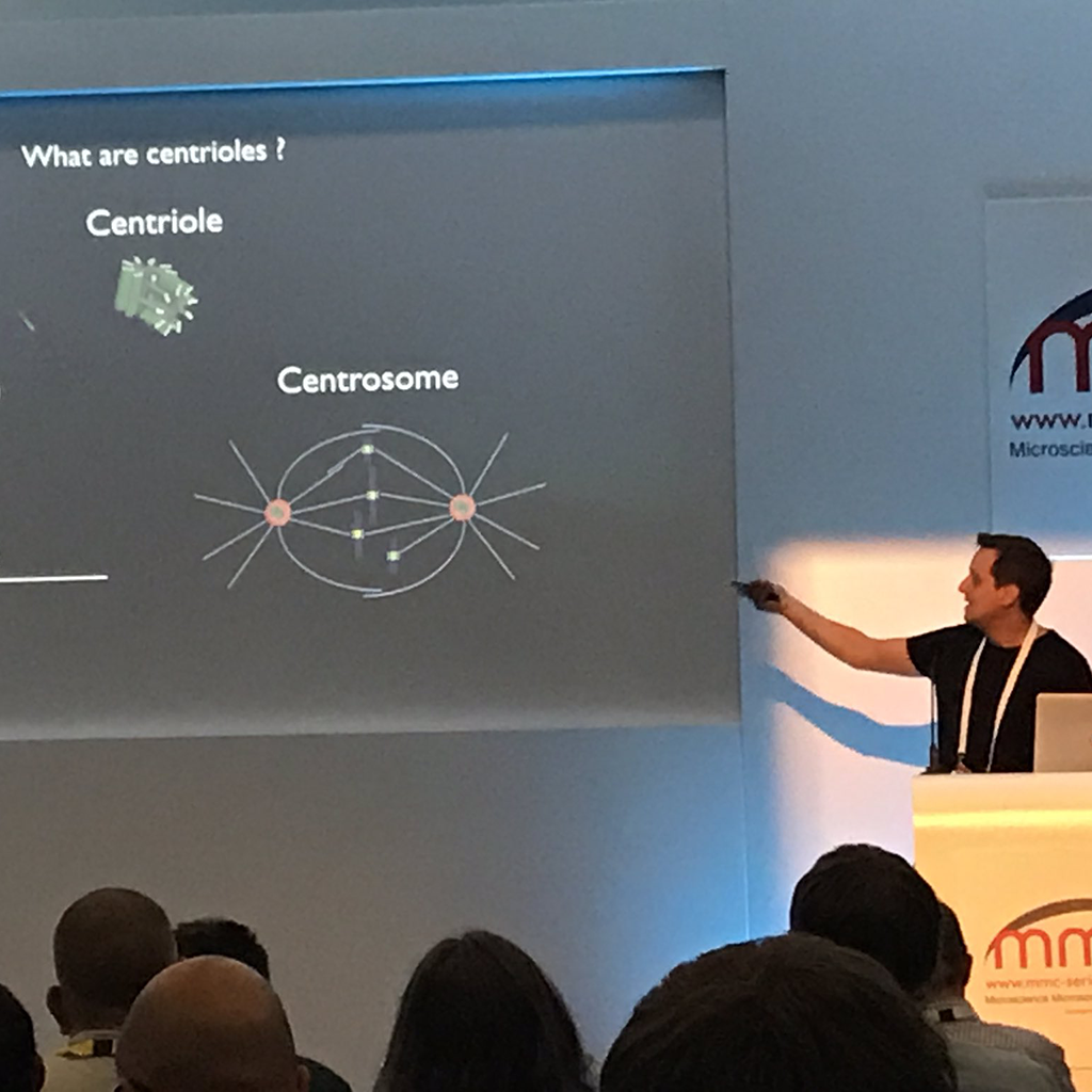
Both Errin (EM) and Alan (LM) presented at the Biennial Microscience Microscopy Conference (“MMC”) in Manchester this July. Errin’s talk “Applying SEM-based Volume and Microanalysis Techniques to the Study of Congenital Dyserythropoietic Anaemia Type-1 (CDA-1)” in the BioApplications: Imaging in Disease session and Alan’s talk “Imaging Dynamic Processes beyond the Diffraction Limit: a Centriole Test Case” in the Frontiers in BioImaging session were both well received. Both talks highlighted some of the exciting imaging in the Dunn School Bioimaging Facility.
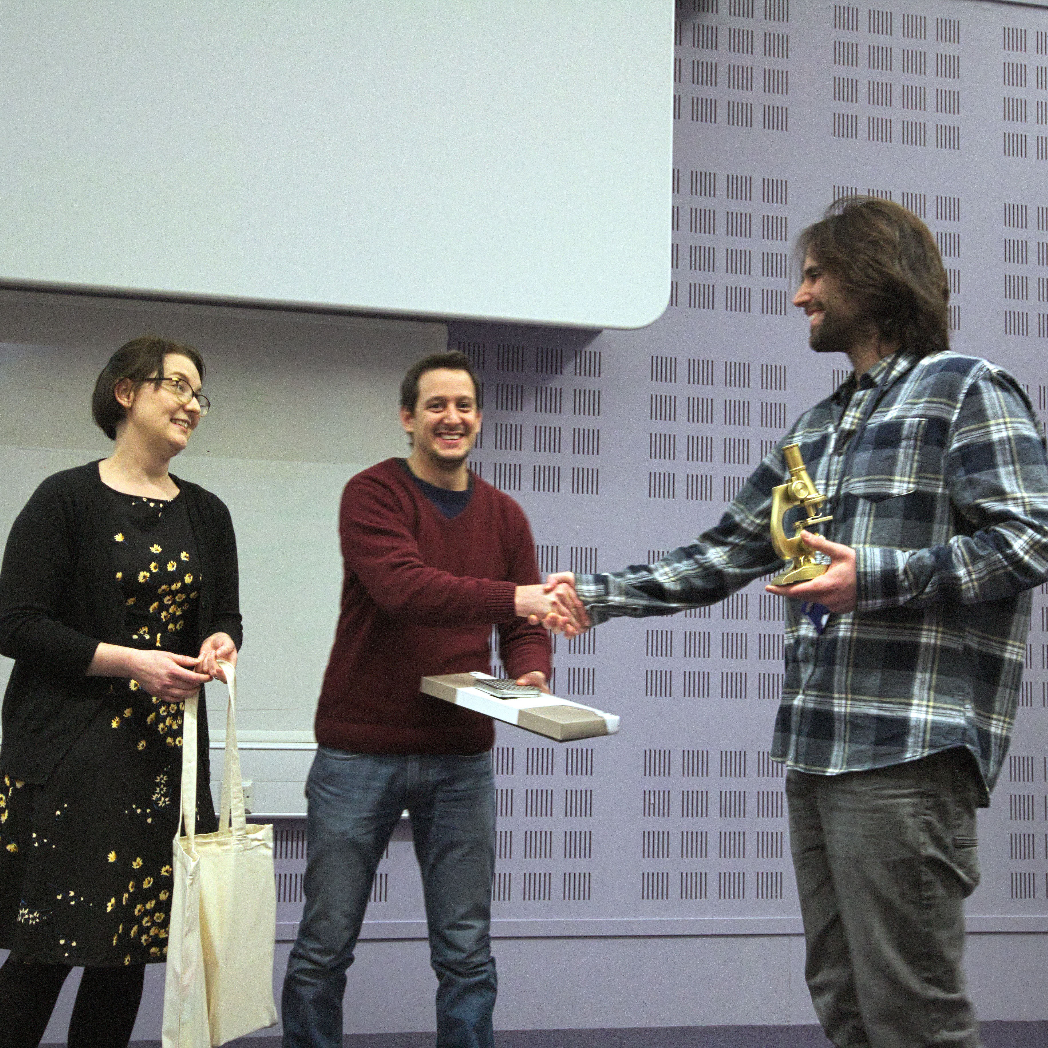
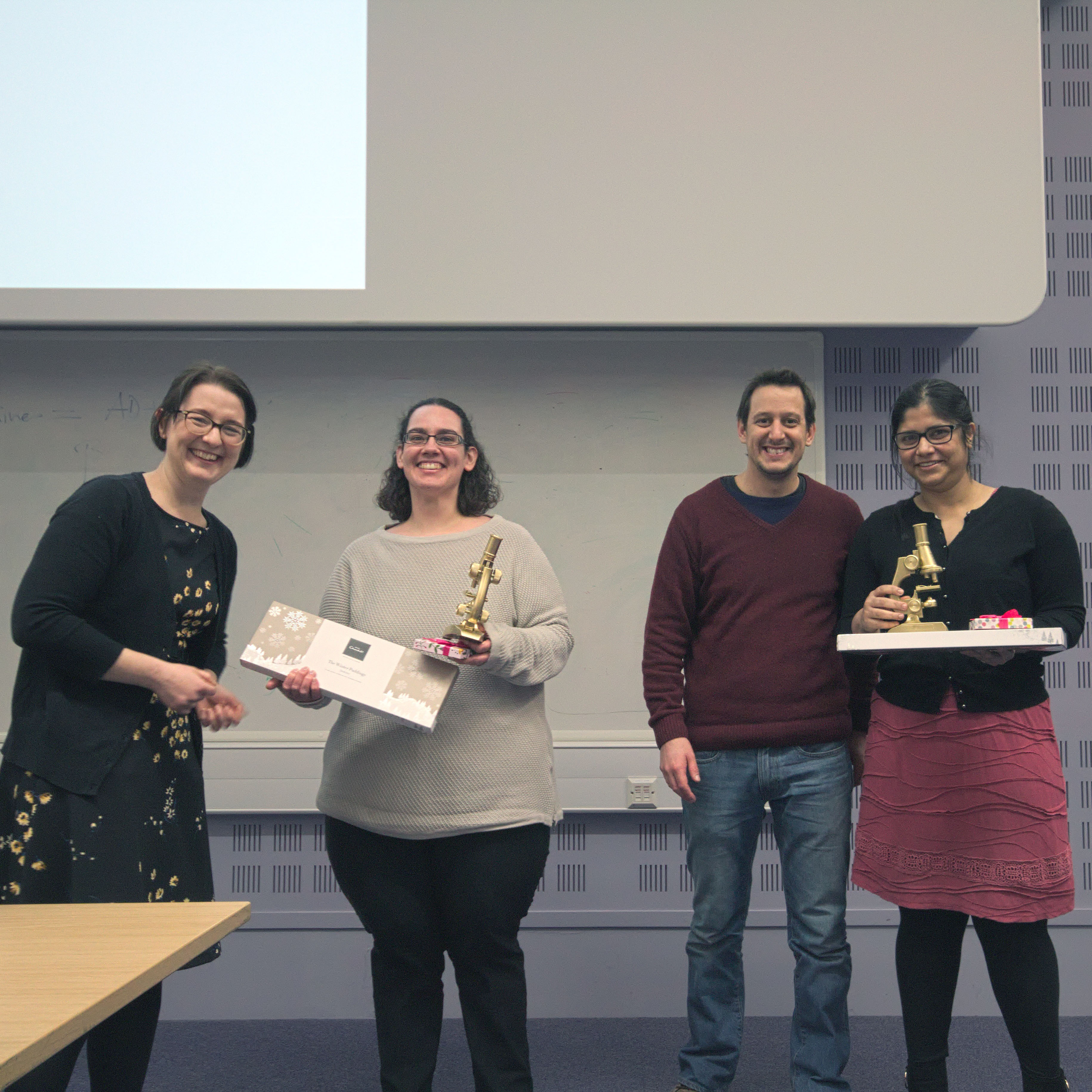
January 2019: Winners of Annual Bioimaging Awards Announced
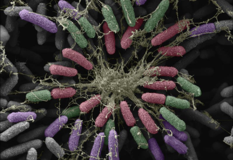
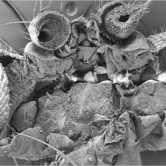
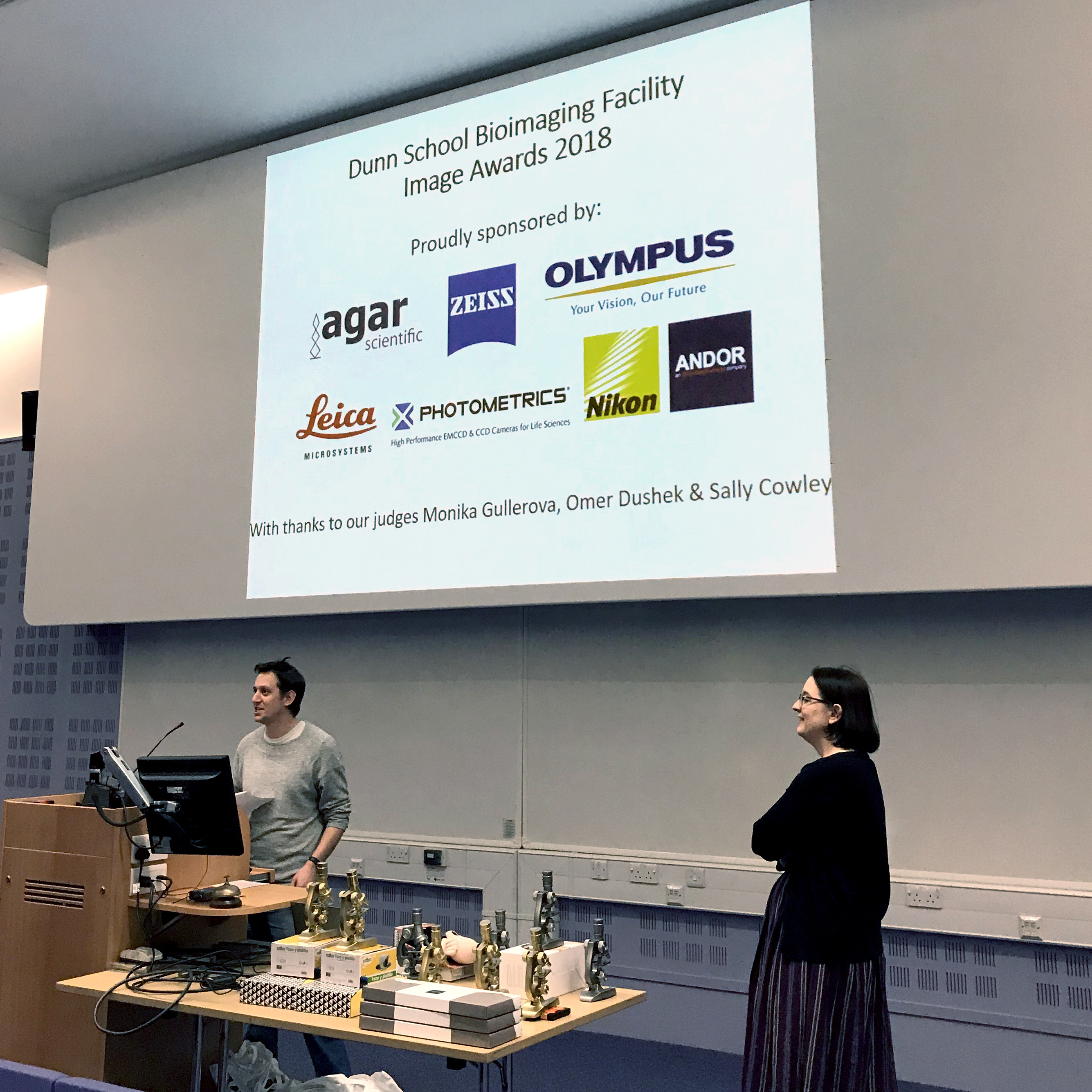
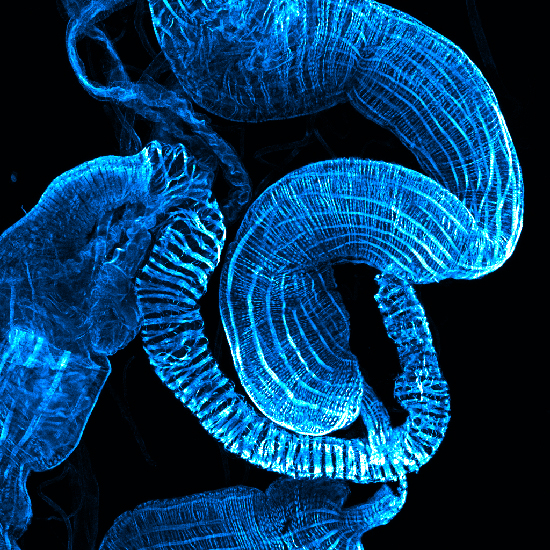
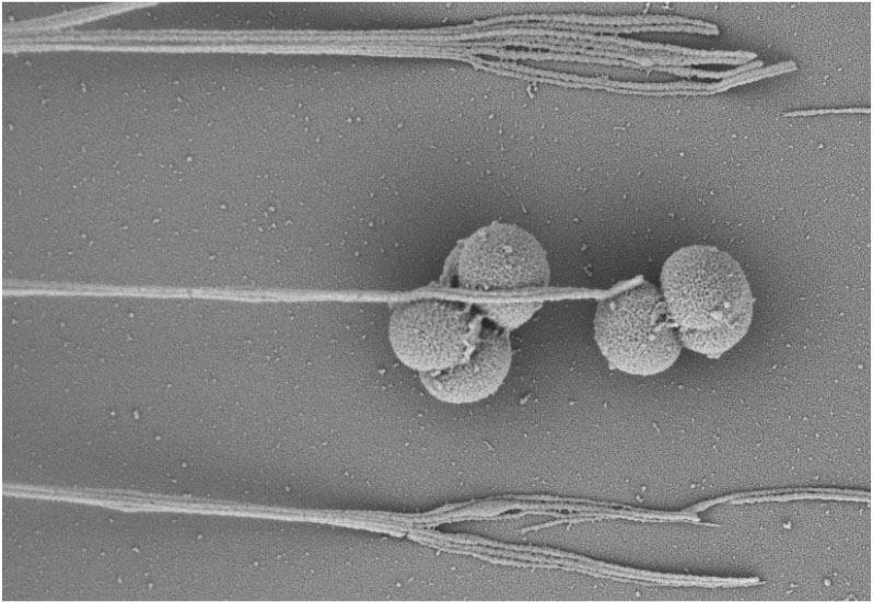
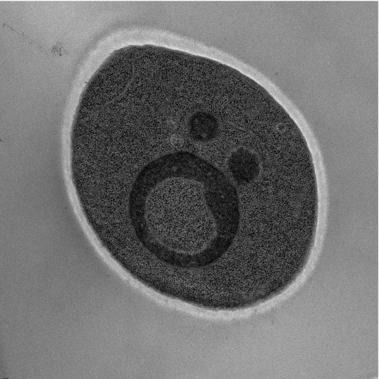
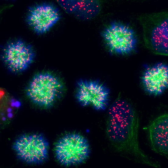
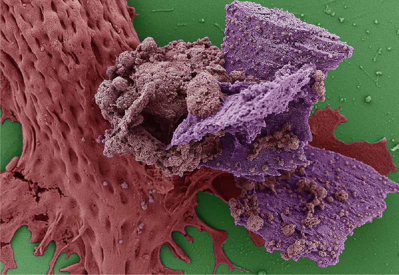
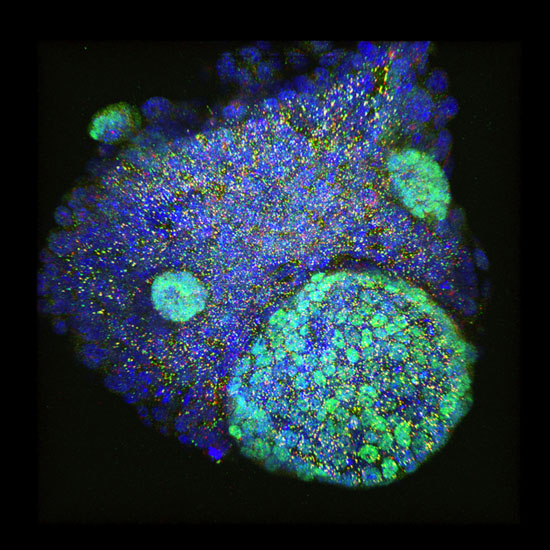
The winners of the Dunn School Bioimaging Facility Awards 2018 were announced at the department’s annual Symposium on the 7th of January 2019. This yearly imaging competition hosted by the Dunn School Bioimaging Facility managers, Dr. Errin Johnson (Electron Microscopy) and Dr. Alan Wainman (Light Microscopy), allows researchers to showcase their most beautiful, interesting and inspiring microscopy work taken on one of the many state-of-the-art departmental facility microscopes.
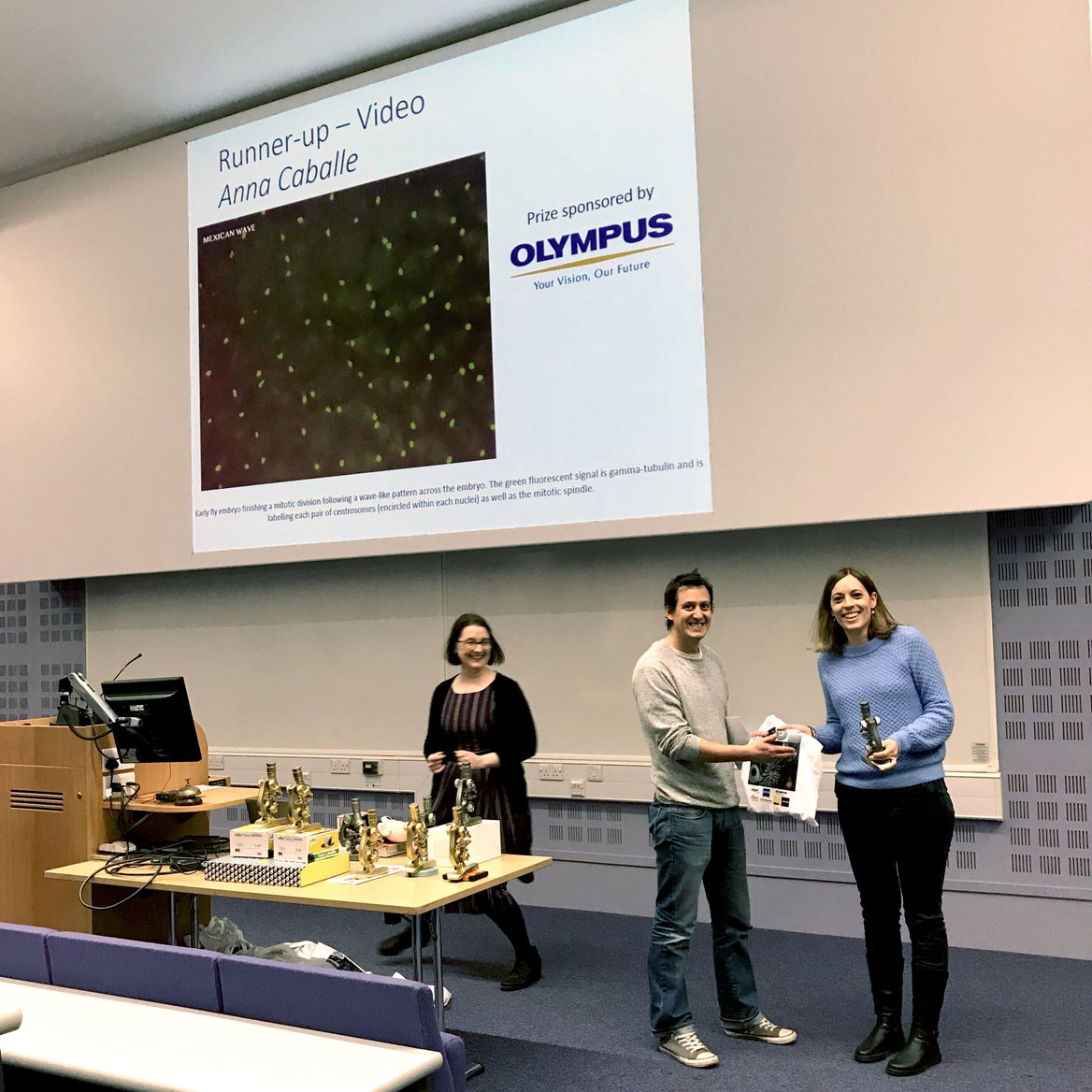
This year’s awards were generously sponsored by Leica Microsystems, Zeiss, Nikon, Olympus, Andor, Photometrics and Agar Scientific. The judging panel, composed of Associate Prof. Omer Dushek, Dr. Sally Cowley and Associate Prof. Monika Gullerova, selected winning and runner-up images in four categories: light microscopy, electron microscopy, humorous microscopy and, a new category, video.
Dr. Wainman said: “2018 has been another great year of imaging in the facility – not least the amazing submissions that were entered in this competition. Many thanks to everyone who participated and congrats to the winners!”
Written by Lisa Gartenmann
January 2018: Winners of Annual Bioimaging Awards Announced

June-July 2017: Through the looking glass- public microscopy exhibition
One of the oldest and most enduring literary connections to Oxford is Alice’s Adventures in Wonderland and its sequel Through the Looking Glass. Alice’s day is now a feature of the city’s calendar, celebrating all things Alice on the first Saturday of July.
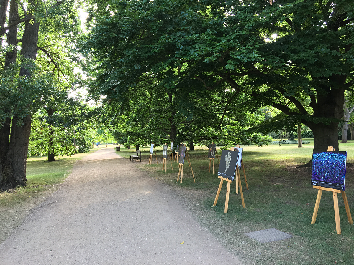
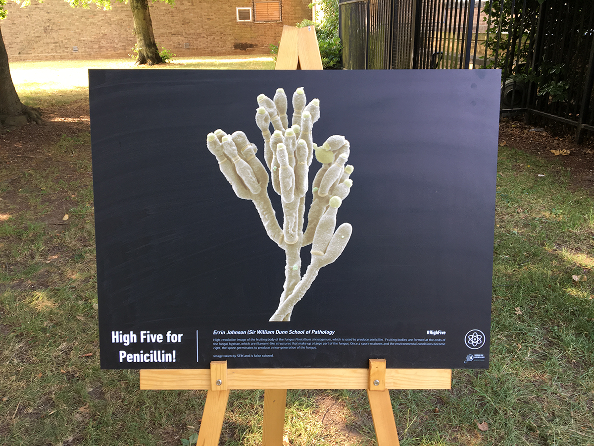
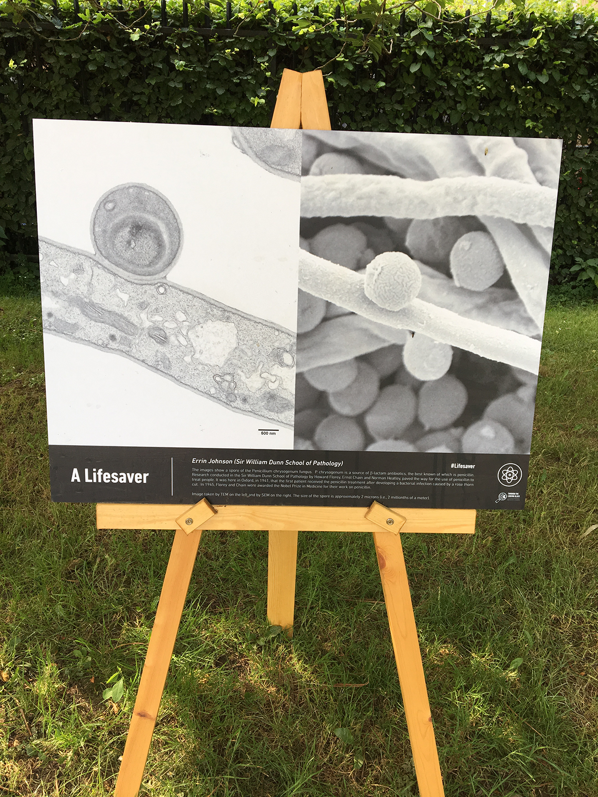
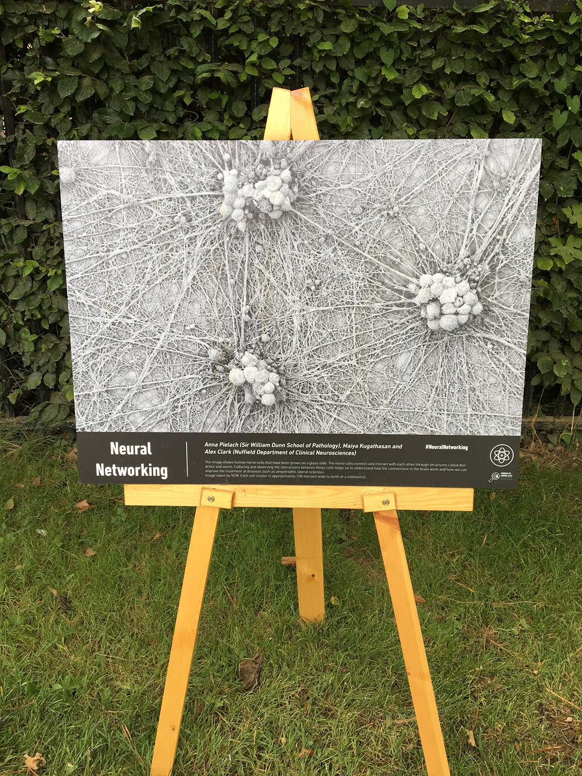
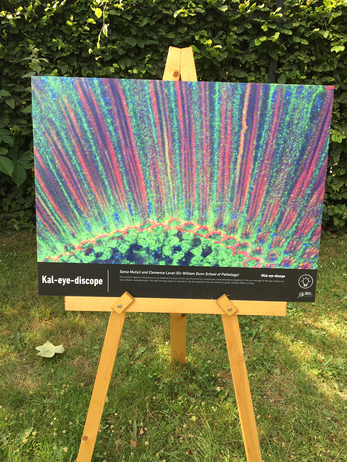
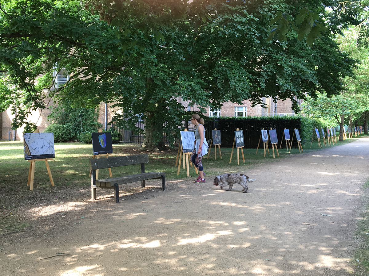
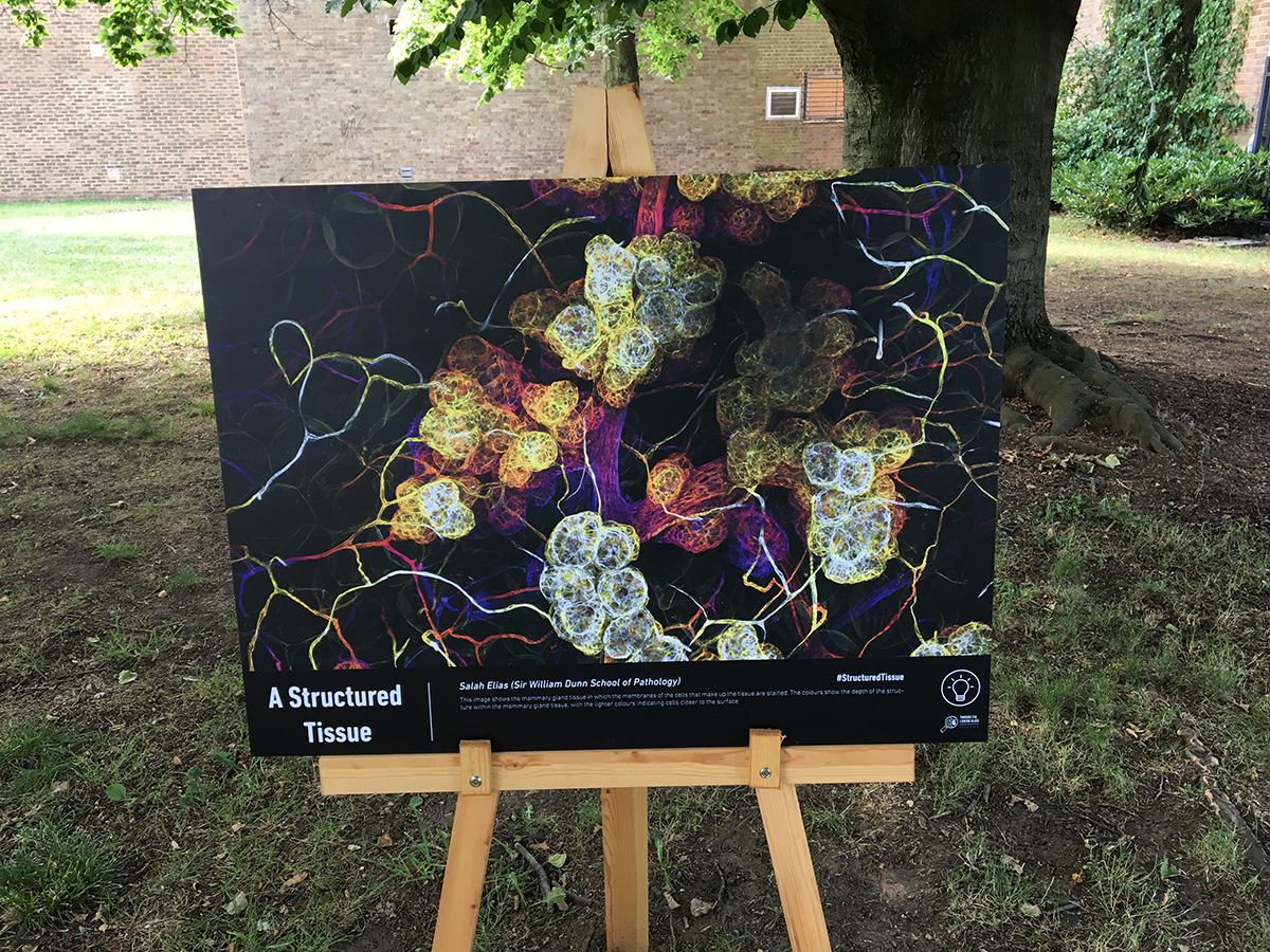
While Oxford is a place of literature, it is also a place of science. Hundreds of scientists use microscopes every day to make small things big and open the door to a world as puzzling and exciting as Wonderland. Oxford houses a range of state of the art microscopes, that allows our scientists to conduct world class research, while also producing beautiful, even artistic, images. To share the beauty of science with the public and celebrate these ‘real life’ looking glasses, the Oxford Branch of the British Science Association organised this year an exhibition in the University Parks that showcased some of the images (and the science behind them). Coinciding with Alice’s day, and lasting from the 12th of June to the 7th of July, 24 beautiful microscopy images, many from the Dunn School Bioimaging facility, were displayed across the University Parks. This project was funded by the EPA Fund. Called "Through the looking glass" we hope that these artistic images may have first surprised the dog walkers, runners and families that regularly visit the park, but also provided a glimpse of the varied and exciting science done at Oxford.
'It seems very pretty,' said [Alice] when she had finished it, 'but it's rather hard to understand!' Hopefully the visitors to park will have appreciated some of beauty of science and maybe understood it a bit better.
March 2017: Research assistant in electron-microscopy
We are seeking a Research Assistant to join the Electron Microscope Facility based in the Sir William Dunn School of Pathology at the University of Oxford.
The successful candidate will assist the EM Facility Manager in two main areas. The first is the day-to-day running of the Facility, which will include preparation of chemical solutions, ordering consumables, management of hazardous waste disposal and general lab maintenance. The second will be to undertake work on service projects, which will involve the preparation and imaging of biological specimens for EM, and also to train users in EM specimen preparation techniques (including ultramicrotomy) and on the electron microscopes. There will also be scope to develop the capabilities of the Facility in advanced EM techniques, which will involve both independent and collaborative work with researchers.
for more details see our job advert
March 2017: Research assistant in cryo-electron microscopy
We are seeking a Research Assistant to join the Central Oxford Structural Molecular Imaging Centre (COSMIC), a newly established cryo-electron microscopy facility in the South Parks Road Science Area at Oxford University. COSMIC is affiliated with the Dunn School EM Facility, together forming a major hub for cutting-edge biological electron microscopy at Oxford.
The successful candidate will assist the EM Facility Manager in two main areas. The first is the day-to-day running of COSMIC, which will include ordering consumables, filling liquid nitrogen dewars and general lab and microscope maintenance. The second will be to train users in negative staining, cryo-sample preparation and on the electron microscopes. There will also be scope to develop the capabilities of the facility in advanced cryo-EM techniques, which will involve both independent and collaborative work with researchers.
for more details see our job advert
March 2017: New website launched
